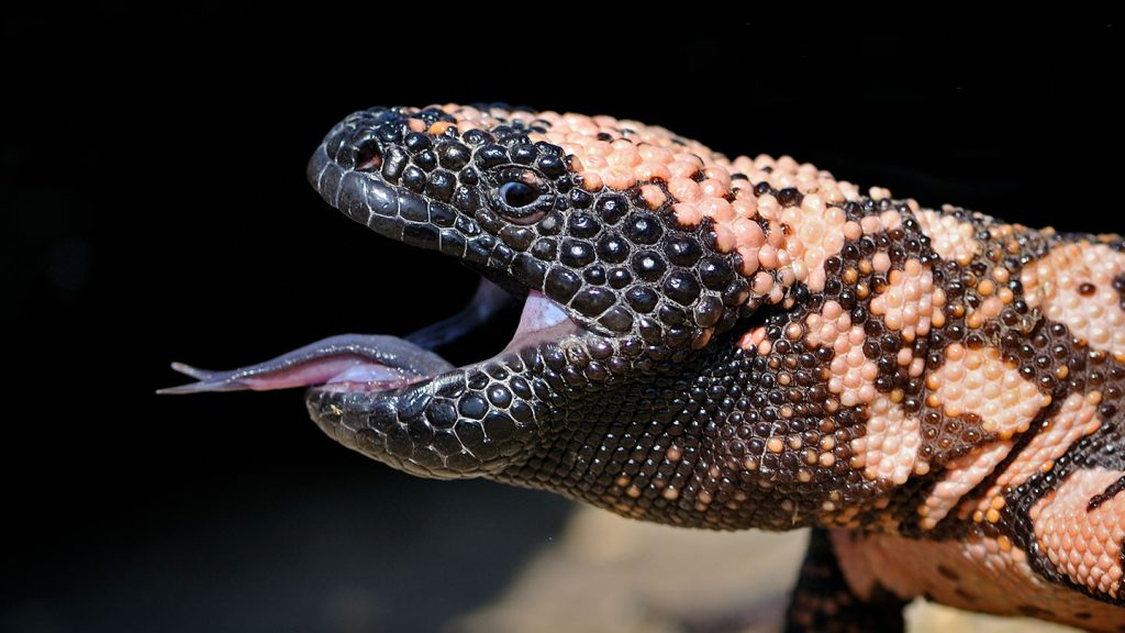
Scientists have found a way to use a tweaked variant of a protein found in Gila monster saliva to make it easier to detect certain tumors, specifically insulinomas, in the pancreas. Insulinomas are benign tumors that produce excessive insulin, leading to low blood sugar levels and fainting spells. The current scanning methods, including CT and MRI scans, have limitations in detecting these small and hidden tumors, making it challenging for surgeons to locate and remove them accurately. The new type of PET scan, using a modified protein called exendin-4, was successful in detecting the tumors in 95 percent of confirmed cases, a significant improvement from the 65 percent success rate of standard PET scans.
The task of producing insulin in the pancreas falls upon specialized cells called beta cells. However, malfunctioning of these cells can lead to the formation of insulinomas, which are rare but debilitating tumors. Current methods of detecting these tumors are ineffective in locating the insulinomas accurately. Surgeons often have to resort to invasive procedures, like slicing the pancreas until the tumor is found, increasing the risk of complications. The use of radioactive molecules injected into the body in PET scans can provide a three-dimensional view of cancerous cells, but may not be efficient in detecting small insulinomas.
Researchers discovered that exendin-4, a protein found in Gila monster saliva, could be used as a radiotracer in PET scans to effectively locate insulinomas due to the high amount of GLP1 receptors present in these tumors. By modifying exendin-4 and adding stabilizing molecules, the new radiotracer showed high radioactivity even at low doses, reducing the risk of side effects like nausea and headaches. In a study involving 69 patients diagnosed with insulinomas, the exendin-4 PET scan successfully detected the tumors in 50 out of 53 confirmed cases, surpassing the standard PET scans and other imaging techniques.
The new exendin-4 PET scan exhibited higher accuracy in detecting insulinomas with minimal background noise and fewer adverse effects on patients. This breakthrough represents a significant improvement in diagnosing these rare tumors and could potentially replace existing imaging methods. Researchers are now focused on disseminating this novel technique to other medical facilities to benefit more patients and help guide surgeons in accurately locating and removing insulinomas. The use of exendin-4 as a radiotracer in PET scans has shown promising results in transforming the diagnosis and treatment of insulinomas, offering hope for patients with these challenging tumors.


