


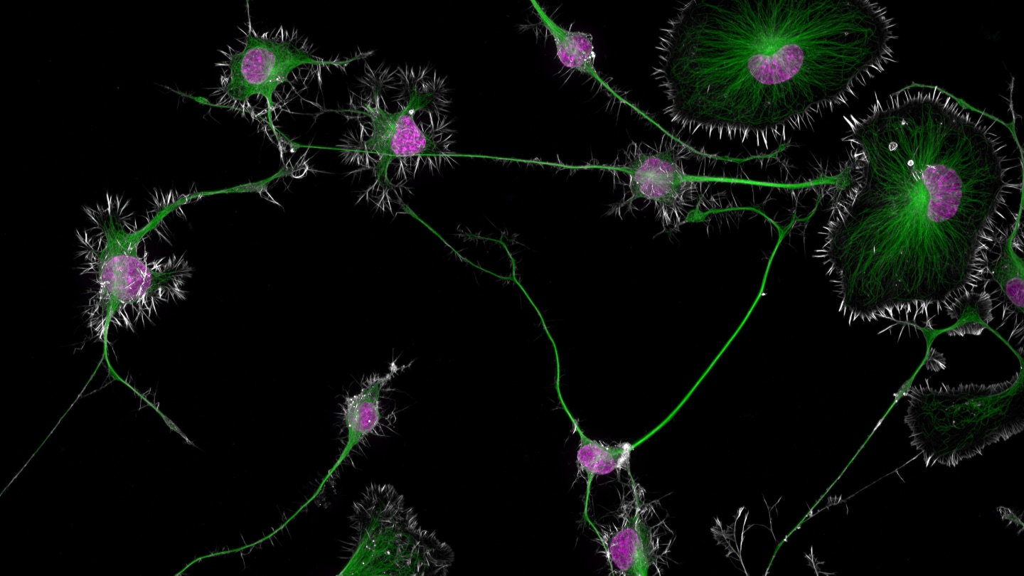
The Nikon Small World photomicrography competition showcases stunning close-up images of microscopic structures. The first-place winner this year is a close-up peek at mouse brain tumor cells captured by neuroscientist Bruno Cisterna. The image reveals the nuclei of tumor cells surrounded by actin proteins and microtubules that transport organelles within cells. Cisterna’s research aims to understand neurodegenerative diseases such as Alzheimer’s and ALS by studying how these cellular structures function. By tracking abnormalities in cellular structures, researchers hope to uncover insights into cell death and neurodegeneration.
Another remarkable image in the competition is an electric arc captured by physicist Marcel Clemens. The photo shows sparks flying between an entomology pin and a dismantled stick gas lighter. The pink or purple glow seen in the image comes from electrically charged atoms losing or gaining electrons. Clemens captured the electrical arcs with long exposure times, allowing viewers to see the intricate patterns of the electricity traveling along the wire. The image highlights the beauty and complexity of electrical phenomena on a macroscopic scale.
Photographer Paweł Błachowicz created a fascinating close-up image of the eyes of a green crab spider, magnified 20 times normal size. The intricate details of the spider’s eyes, resembling a UFO or mutant Kermit the Frog, showcase the remarkable structures found in nature. Crab spiders like Diaea dorsata can change color to blend in with their surroundings, making them fascinating subjects for close-up photography. The photo offers a glimpse into the microscopic world of arachnids and captures the intricate patterns and structures of the spider’s eyes.
A striking image captured by life scientist Kseniia Bondarenko shows Toxoplasma gondii parasites dividing asexually inside a human skin cell. The parasites, shown at 100 times magnification, resemble oval-shaped pairs that look like coffee beans. Bondarenko used a high-resolution microscope and computational methods to image the parasites in three dimensions. The image highlights the intricate structures of the parasites’ inner shells, skeletons, and nuclei. Toxoplasma gondii can cause severe disease in humans, making it an important subject for research and microscopy.
Zoologist Sherif Abdallah Ahmed’s image of an adult red palm weevil showcases the destructive nature of these insects. The weevils are among the most destructive palm pests globally, using their drill-like snouts to bore into palm trees and destroy them. Abdallah’s image captures the intricate details of the insect’s head, with front antennae resembling a boxer’s gloved fists. The image highlights the beauty of the weevil’s features, even as it causes destruction in palm groves. Abdallah’s research aims to combat the spread of red palm weevils and protect palm trees from infestations.
Finally, photographer Daniel Knop captured the opening of a Hibiscus moscheutos anther over a span of 40 minutes. The series of images shows the anther releasing pollen into the air as it opens, showcasing the intricate process of pollination in flowers. Knop’s close-up images offer viewers a glimpse into the natural world’s small details and shed light on the beauty and complexity of plant reproduction. The detailed images of the anther opening highlight the delicate and fascinating process that occurs in nature.


