


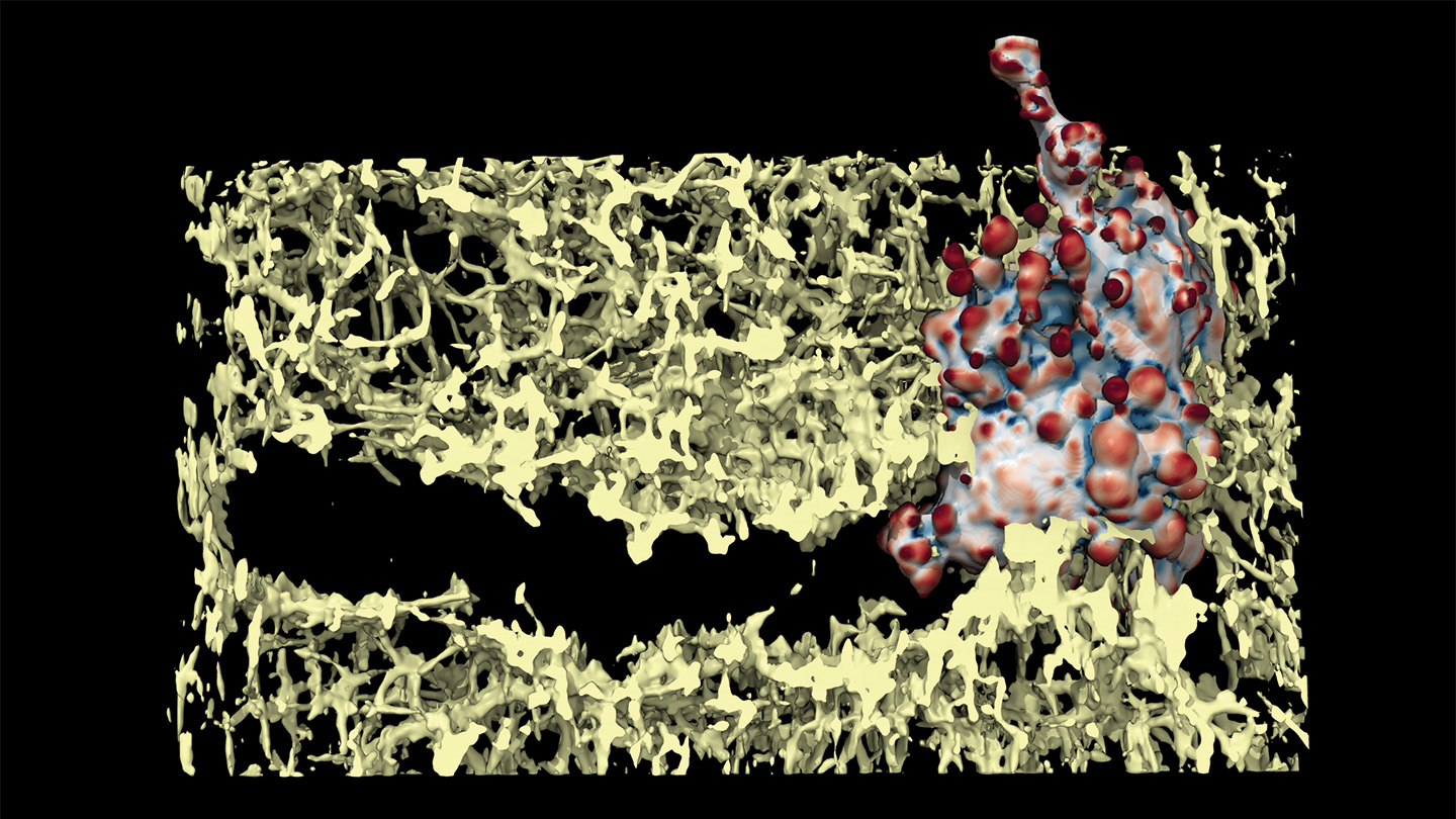
A new study by researchers at the University of Texas Southwestern Medical Center and the University of Minnesota Twin Cities explores how cancer cells are able to move through tissue using bubbles called blebs. The study utilized a new type of microscope that allows for more accurate imaging of cell movement. The researchers found that the blebs formed by cancer cells degrade material, creating a tunnel for the cell to move through. They also observed that the cells are constantly propelled forward through a continuous cycle of bleb formation and interaction with tissue resistance.
The research team found that blebs can form anywhere on the cell, but only those located toward the front of the cell meet a surface. Molecular adhesions on these blebs act as feet, allowing the cell to sense the tunnel surface and signal its direction of movement. The study also revealed that the cells amplify the formation of blebs in areas where they are needed most, with larger blebs concentrated toward the direction of travel. This mechanically driven reinforcement of blebs is a novel aspect of the research and has not been previously appreciated at this level.
The study also identified increased levels of phosphoinositide 3-kinase, an enzyme involved in cell signaling, near the front of the cells. This led the researchers to hypothesize that cells are continuously driven forward by the cycle of bleb formation and interaction with the tissue. The researchers found that blebs extend and retract approximately every 20 seconds, gradually degrading enough material to create a tunnel that the cell can move through. The formation and sensing of resistance from tissue ahead drives the continuous forward movement of the cells.
The development of this new type of microscope that allows for the observation of cell movement without the added pressure from hard surfaces is a significant advancement in the field. Traditionally, microscopes hold samples in place with hard surfaces, which can alter the behavior of cells. By using a soft gel to surround the cells, the researchers were able to observe the bleb formation and movement of cancer cells more accurately. This new method of imaging provides valuable insights into the mechanics of cell movement and opens up new avenues for studying the behavior of cancer cells in tissue.
Overall, the research sheds light on how cancer cells are able to move through tissue using a mechanism involving blebs. This novel finding of mechanically driven reinforcement of blebs at the front of the cell is a significant contribution to our understanding of cell movement. The continuous cycle of bleb formation and sensing resistance from tissue ahead propels the cells forward, allowing them to navigate through the tissue. Further research in this area could lead to new insights into the behavior of cancer cells and potential strategies for targeting their movement in therapeutic interventions.
Trusting News has partnered with Science News to collect feedback on the potential use of AI in journalism. Although Science News currently does not publish content produced by generative AI, they are interested in hearing views on how AI could be used responsibly in journalism. A short 10-question survey is available for readers to share their thoughts on how Science News could make use of AI in a beneficial and ethical manner.


